This patient had a significant vertical ocular motility disturbance.
Best imaging for orbital floor and maxillary fracture.
Mid face le fort fractures.
A retrospective study by bartoli et al of 301 orbital floor fractures found the most common symptom to be hypesthesia extending through the region of the maxillary nerve 32 9 of patients.
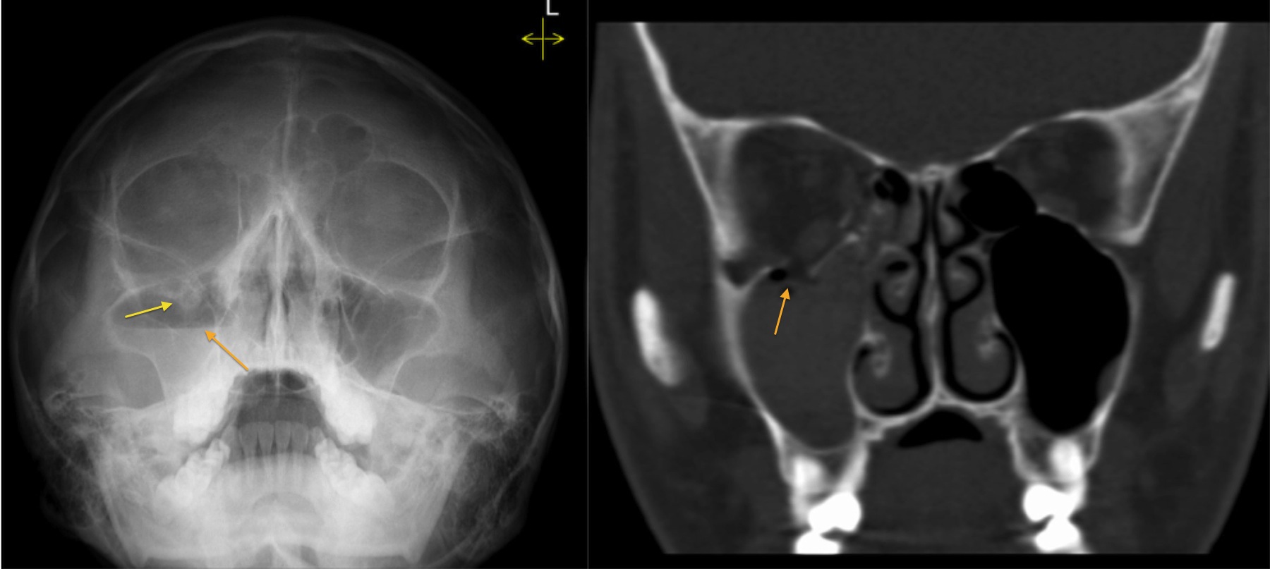
Waters view best displays inferior orbital rims nasoethmoidal bones and maxillary sinuses.
Inferior blowout fractures are the most common.
The usual mechanism is a blow to the eye with the forces being transmitted by the soft tissues of the orbit downward to the thin floor of the orbit.
Blunt force trauma tends to cause fractures along three lines of weakness in the mid face.
This is a rim fracture that extends into the lower socket.
Getting hit with a baseball or a fist often causes a orbital blowout fracture.
If the patient is upright when the film is taken an air fluid level can often be seen in the maxillary sinus which may indicate fracture of the maxillary sinus orbital floor.
The orbit is one of a pair of bony cavities each housing the globe and associated structures.
Zygomatic sphenoid maxillary frontal lacrimal palatine and ethmoid.
Fractures of the orbit may be seen in different scenarios of direct and indirect trauma to the globe orbital facial or cranial bones.
Orbital blowout fractures seen in the emergency setting commonly occur after trauma.
The orbit is formed by 7 bones.
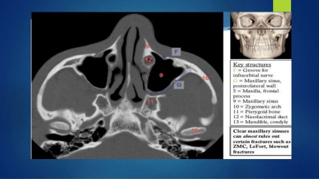
A study by huang et al indicated that in patients with head trauma lack of maxillary hemosinus on conventional head ct scanning predicts the absence of orbital floor fracture the negative.
Orbital floor fractures may result when a blunt object which is of equal or greater diameter than the orbital aperture strikes the eye or on the cheek 1.
Orbital floor fracture also known as blowout fracture of the orbit eye socket.
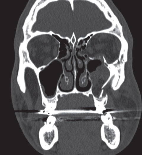
Orbital fat prolapses into the maxillary sinus and may be joined by prolapse of the inferior rectus muscle.
The floor is usually the path of least resistance and fractures downward into the maxillary sinus.
Orbital floor fracture with significant soft tissue entrapment a so called trapdoor fracture.
Maxillary bones upper jaw.
Inferior floor medial wall lamina papyracea superior roof lateral wall.
Blowout fractures can occur through one or more of the orbital walls.
Direct orbital floor fracture.
Another common fracture is the orbital floor fracture or blowout fracture.